At a glance
- Spores and mycotoxins are different things. A mold spore is a biological structure. A mycotoxin is a chemical that certain molds can produce. Testing one does not automatically answer questions about the other [1, 2, 3].
- Color is not a diagnosis. A dark patch cannot be identified as Stachybotrys just by its color, and CDC advises addressing indoor mold regardless of type [2, 6, 7].
- Much of the strongest human mycotoxin evidence concerns eating contaminated food, not breathing household air [4, 5].
- Start with moisture, not a kit. If mold is already visible, EPA says sampling is usually unnecessary before addressing it [1, 2, 8].
- Detection is not the same as disease. Low-level dietary exposure can put mycotoxins in healthy people’s urine, and disease-predictive urine levels have not been established [3, 5, 11].
- Functional and integrative practitioners use broader testing as clues to an exposure hypothesis. The published research behind it is observational and does not validate the panels for individual diagnosis [13, 14].
Finding mold at home can turn a practical problem into a frightening set of questions. Are you breathing spores or toxins? Does a dark patch mean “toxic black mold”? Would a urine panel explain why you feel unwell—or would an air sample tell you whether your home is safe?
The terms get mixed together, but they describe different things. A mold spore is a biological structure. A mycotoxin is a chemical that certain molds can produce. A damp building is an environment where mold and other moisture-related problems can develop. Testing one of these does not automatically answer questions about the others. [1, 2, 3]
You can take your symptoms seriously and address water damage without assuming that every symptom is mycotoxin poisoning. This guide separates established knowledge from proposed explanations, including the functional and integrative approaches many readers encounter.
Educational information, not a diagnosis or treatment plan. A building assessment and a medical evaluation answer different questions. Neither should have to wait for a perfect explanation of the other.
What is mold?
Mold is a type of fungus. Instead of growing as a plant with leaves and roots, it grows as tiny, branching threads called hyphae. A connected network of these threads is called a mycelium. Individual threads are microscopic; the patch we notice on a surface is a colony of fungal growth, not a single oversized spore. Growth can also be hidden behind building materials, so the visible surface is not always the whole picture. [1, 28, 29]
In nature, this is useful work. Molds help break down dead leaves, wood and other organic material. Fungi release enzymes that digest material around them, then absorb the nutrients released. The same ability that helps recycle a fallen leaf can damage paper or wood when growth develops inside a building. Mold is therefore a normal part of the outdoor environment, but active growth on indoor materials is a different situation. [28, 29]
To grow, mold needs moisture and a usable food source. Buildings offer plenty of possible food: wood, paper, fabrics and even dust can supply nutrients. A flood is not required; a slow leak or condensation on a cold surface can provide moisture. Different molds favor different conditions, so there is no single growth pattern that fits every building. Controlling moisture matters because the nutrients are often already there. [28, 29]
It helps to separate the growing organism from what it releases. The branching network is the mold's growing body. Spores help it spread; broken threads contribute fungal fragments; and some molds, under certain conditions, produce mycotoxins. Microbial volatile organic compounds, or MVOCs, are another category of metabolic products discussed below. These are related parts of the story, not interchangeable names for the same exposure. First, let's look at the spores. [28, 29]
What are mold spores?
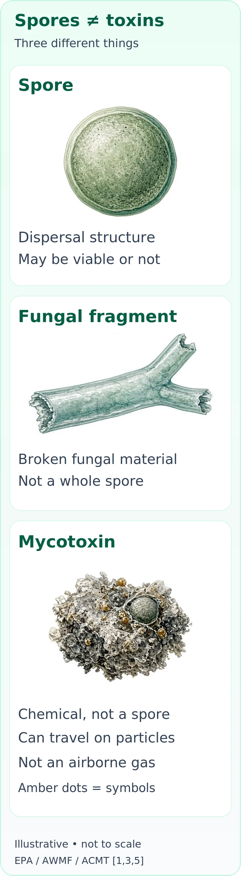
Molds are fungi. They grow as networks of microscopic filaments and reproduce through tiny spores that can move through indoor and outdoor air. A spore is a dispersal structure; “spore” is not another word for “toxin.” When a viable spore lands on a suitable damp surface, it may start new growth. Spores already occur in normal air and house dust, so finding some does not, by itself, establish an indoor infestation. [1, 2, 3]
“Viable” means capable of growing under suitable conditions. A nonviable spore cannot do that, but it may still contain material that matters to someone with mold allergy. Fungal growth can also release fragments of its filaments and other cell material. Those fragments are not whole spores, and a test counting spores does not describe every component of the exposure. [1, 3]
This explains why killing mold and removing mold are not the same job. EPA notes that dead mold can still cause allergic reactions. Fixing moisture and appropriately removing contaminated material matter more than trying to sterilize a room while leaving the contamination in place. This is not a recommendation to disturb hidden mold yourself; opening a contaminated wall can release material and may require experienced professional help. [1]
What are mycotoxins?
Mycotoxins are potentially harmful chemicals produced by certain fungi as secondary metabolites—products of fungal metabolism, rather than the spores or growing filaments themselves. Production depends on the species, its genetics or strain, and growing conditions such as the material, moisture and nutrient environment. A species capable of making a toxin does not necessarily make it in every location. [3, 5]
“Mycotoxin” also names a large family, not one substance with one effect. WHO discusses aflatoxins, ochratoxin A, fumonisins and several other groups in food safety. Different toxins have different effects, and risk depends on the amount reaching a person, the exposure route and duration, and individual susceptibility. A substance being hazardous does not tell you the dose someone actually received. [3, 4, 5]
Much of the strongest human evidence concerns eating contaminated food, not breathing household air. For example, aflatoxin contamination is an established food-safety concern, including liver injury and cancer risk under relevant exposure conditions. That evidence is important, but it cannot simply be transferred to a patch of household mold without establishing the substance, pathway and dose involved. [4, 5]
Aflatoxins: a family with well-established food-related risks
Aflatoxins are one important branch of the mycotoxin family. Certain Aspergillus fungi, particularly A. flavus and A. parasiticus, can produce them. Their presence depends on the fungus and growing or storage conditions—not simply on finding the word Aspergillus on a building report. The same distinction between a producer and its actual chemical output applies here as elsewhere in mold testing. [4, 5, 21]
The four major types are aflatoxins B1, B2, G1 and G2. These are names of chemicals, not four different mold species. Another name, aflatoxin M1, describes a metabolite of B1; it is relevant to food safety because it can occur in milk when animals consume contaminated feed. A laboratory's list of analytes should therefore be read as a list of particular substances, not as a direct inventory of fungi in someone's home. [4, 21]
The established health concern is substantial but specific. Large amounts in contaminated food can cause acute aflatoxicosis, with potentially life-threatening liver damage. Longer-term exposure is linked to liver cancer in humans. IARC's current classification lists aflatoxins as Group 1: carcinogenic to humans. This identifies a cancer hazard; it is not a diagnosis, a prediction that an exposed individual will develop cancer, or a numerical threshold for household air. The strongest human evidence has largely concerned dietary exposure. [4, 21, 22]
There is a biological explanation for the liver concern. The U.S. National Toxicology Program describes how enzymes can convert B1 into a reactive form that binds to DNA. It also describes glutathione-dependent processing of that reactive form. This helps explain why environmental and functional clinicians pay attention to metabolism and protective pathways. However, the existence of a pathway does not establish that a glutathione supplement prevents illness or that a urine result selects a treatment. Those are separate clinical questions. [14, 21]
Food contamination can occur in crops such as corn, peanuts and other nuts before harvest or during storage. This does not mean every serving is contaminated, or that eliminating whole food groups is the answer. WHO emphasizes food monitoring, appropriate storage and dietary diversity. Occupational exposure can also occur through dust from handling contaminated crops or feed; that is a different exposure setting from ordinary residential air. Food-safety limits address food and cannot simply be relabeled as home-air limits. [4, 21]
For a functional assessment, an aflatoxin-related urine finding is a prompt to clarify the analyte, collection conditions, dietary and occupational history, and the clinical question. Some aflatoxin biomarkers have useful research applications. That does not make a commercial panel proof of a household source or proof that aflatoxin explains a person's chronic symptoms. A building investigation and a medical evaluation still need their own evidence. [5, 18, 21]
Ochratoxin A: kidneys, persistence and what a panel cannot tell you
Ochratoxin A, usually shortened to OTA, is another mycotoxin—not a type of spore and not a musty-smelling gas. Certain species of Aspergillus and Penicillium can produce it. Food contamination is a well-recognized exposure pathway, including contamination involving cereals, dried vine fruits, coffee and some other stored foods. These examples explain why diet matters when interpreting biomonitoring; they are not a shopping blacklist or evidence that a particular product is unsafe. [4, 23]
The kidneys are a major focus of OTA toxicology. Studies in animals show kidney injury, and rodent studies show kidney tumors. Human findings are harder to interpret. WHO contrasts clear animal evidence of kidney toxicity and kidney cancer with uncertainty about the corresponding human associations. EFSA's assessment explains why available observational human studies do not establish those causal links. A positive urine result is not equivalent to kidney damage, and fatigue or other nonspecific symptoms cannot supply that missing diagnosis. [4, 23]
The cancer classification is different from that for aflatoxins: IARC currently lists ochratoxin A as Group 2B: possibly carcinogenic to humans, not Group 1. EFSA's 2020 food assessment also recognized uncertainty about how OTA produces genetic damage and kidney tumors, and used a margin-of-exposure approach rather than its former tolerable weekly intake. These are food-risk assessment tools, not clinical urine cutoffs for people worried about a building. [22, 23]
OTA's behavior in the body helps explain the interest in persistence. EFSA describes extensive binding to blood proteins, particularly albumin, along with relatively slow elimination and limited metabolism. Binding influences how the chemical circulates and is cleared; the amount measured in blood or urine is not simply the amount entering the body that day. Human kinetic data are limited, so there is no reliable personal “clearance clock” to calculate from a single panel. [18, 23]
That gives an integrative clinician a reasonable set of questions: What exactly was measured? Is it the parent toxin or a related compound? What do diet, collection timing and kidney function contribute to interpretation? EFSA found that dietary exposure can be reflected in blood and urine measurements, but did not identify reliable biomarkers of OTA-specific effects on kidney function. An exposure measurement and an organ-injury assessment are different jobs. If kidney disease is suspected, conventional clinical assessment matters—not an assumption that the panel has already diagnosed it. [18, 23]
Functional and environmental-medicine discussions also draw on experimental OTA work involving oxidative stress, mitochondrial function, immune signaling and intestinal barriers. Those studies help generate hypotheses about biological responses. As discussed later in this article, much of that evidence comes from cells or animals, with doses and routes that cannot be assumed to reproduce residential human exposure. It does not establish a universal sequence from a damp wall to multisystem illness, or automatically justify an antifungal or “detox” treatment. [14, 19, 23]
Macrocyclic trichothecenes: a family, not one toxin
Macrocyclic trichothecenes are a subgroup of the larger trichothecene family, often called Type D trichothecenes. “Macrocyclic” refers to an additional large ring in their chemical structure; it does not mean a larger mold spore. Roridins, verrucarins and satratoxins belong here. These are related chemicals, not interchangeable names for a single substance. Examples include roridin E, roridin L-2, verrucarin A, satratoxin G and satratoxin H. The list is representative rather than exhaustive. [30, 31, 32]
Certain Stachybotrys fungi and fungi described in the literature as Myrothecium can produce members of this group. Even within Stachybotrys chartarum, different chemical-producing types—called chemotypes—can look alike while producing different mixtures. The macrocyclic-trichothecene-producing chemotype is not a description of every strain or every colony. Genetics, the material being colonized and growing conditions matter; color or a species name alone does not establish the amount of toxin produced. [5, 30, 31]
Why do toxicologists study them? One important experimental effect is interference with protein synthesis, the process cells use to build proteins. A classic primary study found that verrucarin A inhibited the initiation of protein-chain production in cultured human cells and yeast preparations. It also showed why the broader family should not be treated as mechanistically identical: different trichothecenes affected different stages of the process. This is meaningful cellular toxicology, not evidence that every person near a moldy wall is experiencing that effect. [32]
There is also direct building research—not just fungal cultures. A 2016 case study of a water-damaged building analyzed fungal material and settled dust and detected roridin E in a dust sample. The researchers also identified other metabolites in contaminated wall material and documented differing chemical profiles. That establishes that such compounds can occur in a real building; it does not turn a dust concentration into an individual's inhaled dose, a diagnosis, or a health-based cleanup threshold. The study was an analytical case study, not a trial proving the cause of occupants' symptoms. [3, 31]
For someone working with an environmental or integrative clinician, the useful distinction is a plausible mechanism versus a demonstrated personal exposure-and-effect chain. Evidence that a toxin can disrupt a cell process is a reason to investigate carefully, not a shortcut past identifying the material, route and relevant exposure. Equally, uncertainty about the precise toxin does not make visible dampness or growth acceptable: moisture correction and appropriate removal remain practical priorities. [1, 3, 5]
Gliotoxin and “derivatives”: why the chemical name matters
Gliotoxin is an individual fungal secondary metabolite; “gliotoxin derivative” is not a uniquely identifying chemical name. Gliotoxin is associated especially with certain Aspergillus fumigatus isolates, but production varies across fungi and experimental conditions. It should not be confused with the macrocyclic trichothecenes, and finding Aspergillus in a building does not by itself establish gliotoxin production. [5, 33]
One specifically named derivative is bis(methylthio)gliotoxin, abbreviated bmGT. Researchers have described its formation when fungi chemically modify gliotoxin by methylation. It is not simply another spelling for the parent toxin, and the 2018 study describes it as an inactive derivative involved in regulation of gliotoxin production. That does not establish that every substance described generically as a “derivative” has the same identity or activity. If a report uses a broad label, ask which exact compound was measured, in what specimen and by what method; do not assume that it means bmGT. [33]
Immune effects: what the laboratory evidence actually shows
Gliotoxin interests researchers partly because of effects on immune-cell behavior and signaling. For example, a 2005 experiment exposed a laboratory-grown human monocytic cell line—an immune-cell model—to gliotoxin and measured signaling proteins including IL-10, IL-6 and TNF-alpha, along with cell survival. The responses depended on concentration and were not simply “all inflammation goes up”: different signals changed differently, and reduced cell viability mattered. The authors explicitly said the clinical implications needed verification in human studies. These were cells in culture, not patients being treated for household exposure. [35]
This gives a concrete basis for an integrative discussion about immune signaling while also defining its boundary. A clinician can ask whether an exposure history and a person's medical findings fit a concern worth evaluating. A laboratory mechanism alone cannot demonstrate that gliotoxin caused someone's recurrent infections, fatigue, mast-cell symptoms or immune dysfunction. Those possibilities require an appropriate clinical assessment rather than a diagnosis inferred from the chemical's reputation. [3, 5, 35]
Human infection research is a different clinical setting
Gliotoxin-related measurements have also been investigated in invasive aspergillosis, where an infection is being evaluated—not merely a damp apartment. A 2018 primary study compared production in fungal isolates with bmGT in blood serum from patients with probable or proven invasive aspergillosis and reported findings supporting further biomarker research. A later prospective study in 18 patients with proven or probable invasive pulmonary aspergillosis found poor performance: no serum gliotoxin was detected, and serum bmGT was detected in only one patient. The later authors did not recommend those measurements for diagnosing that infection. [33, 34]
The contrast matters more than choosing the most reassuring or alarming paper. Biomarker performance depends on the population, specimen and method, and an infection study does not validate an unspecified urine “derivative” result as a test for illness from a building. Conversely, a negative toxin measurement cannot independently exclude a serious fungal infection. If infection is a concern—particularly in someone at increased risk—the answer is a clinician-directed infection assessment, not self-prescribed antifungals or a detox response to a label. [3, 33, 34]
Zearalenone: an estrogen-active food-associated mycotoxin
Zearalenone, often abbreviated ZEN or ZEA, is a nonsteroidal mycotoxin produced by certain Fusarium fungi. “Nonsteroidal” means it is not an estrogen hormone, even though it can have estrogen-like biological activity. Its best-established exposure context is contaminated crops and food or feed, especially cereal grains. That is a different starting point from assuming that a damp indoor wall is the source. As with the other toxins discussed here, a potentially producing fungus and a measured chemical are different findings. [5, 36, 37]
Its distinctive toxicological interest is estrogen-receptor activity. Estrogen receptors are proteins through which estrogen signals to cells; zearalenone and some related metabolites can interact with that system. Experimental work and animal studies show effects relevant to reproductive tissues and function, with pigs particularly sensitive in the assessments discussed by WHO and EFSA. The mechanism is more specific than a vague claim that the toxin “disrupts every hormone,” and the effect depends on the compound and exposure. [36, 37]
The parent compound is only part of the chemistry. Zearalenone can be converted into related substances, including alpha- and beta-zearalenol, and further conjugated forms. These names are not all equivalent to one another: EFSA notes substantial differences in estrogenic activity among modified forms. Detecting a metabolite can inform exposure research, but its interpretation requires knowing what the measurement actually includes and how it relates to the parent compound. [18, 36, 37]
Evidence and limits: the animal reproductive findings are important and support food-safety assessment. The WHO evaluation also discusses human observations, including investigations of early breast development, but these observations do not provide a simple, established causal explanation for an individual's hormonal symptoms. They do not establish that ordinary residential inhalation causes the same outcomes seen in animal feeding studies. Food-risk guidance values are not household-air standards or clinical thresholds for a positive urine test. [3, 18, 36, 37]
For an integrative evaluation, the productive question is whether a plausible exposure history deserves consideration alongside a standard assessment of the hormonal concern, rather than replacing that assessment. A positive measurement does not establish infertility, explain every menstrual symptom, or locate a building source. Nor does this food-contamination context call for eliminating all grains or undertaking an antifungal or hormone-altering regimen on your own. The broader testing principles below—exact analyte, specimen, exposure context and a decision the result can genuinely inform—apply here as well. [3, 4, 18, 36]
Microbial volatile organic compounds: the musty-smell question
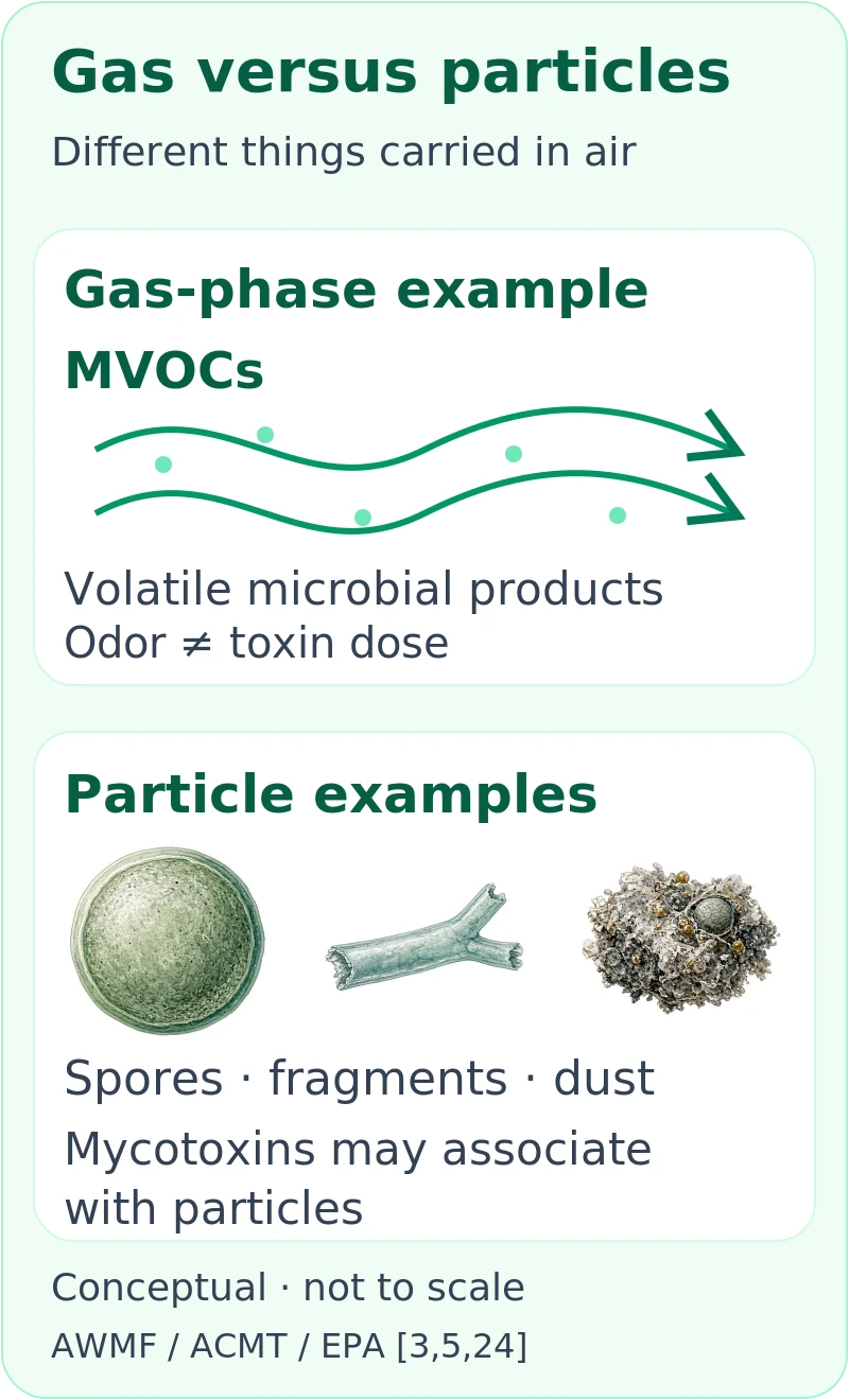
A damp building can involve another category of chemicals: microbial volatile organic compounds, abbreviated MVOCs. Fungi and bacteria produce these volatile metabolic products. “Volatile” means they can enter the air as gases, unlike the particle-associated mycotoxins discussed above. The familiar musty odor can involve a mixture of these compounds rather than a cloud of spores that you are somehow smelling directly. [3, 24]
The AWMF guideline gives examples such as 1-octen-3-ol, with a mushroom-like odor, and geosmin, with an earthy odor. A mixture can smell quite different from any one ingredient. Importantly, chemicals described as MVOCs are not necessarily exclusive microbial markers: some also have sources such as cooking or tobacco smoke, and indoor materials can contribute to the wider VOC mixture. A chemical name on an air report therefore does not automatically identify hidden mold, a particular species or one source. [2, 3]
This is why a musty smell is useful as an investigation clue, not a toxin-concentration meter. EPA advises investigating moldy odors for a building problem. Smell cannot tell you the concentration of aflatoxin or OTA, how much reached your body, or whether a location is safe for a particular person. It is also a mistake to dismiss someone's experience because no toxin has been demonstrated: a troublesome odor and a moisture problem can deserve attention before the chemistry is fully resolved. [1, 3, 24]
People may describe irritation, headache, nausea or feeling unwell around these odors. EPA notes links with symptoms including nasal irritation and headache, while emphasizing that health effects from non-occupational indoor MVOC exposure remain poorly understood. An association does not identify which chemical or mixture caused an individual's symptoms. It also does not demonstrate that the odor represents systemic mycotoxin poisoning. The useful clinical approach is to document the experience, assess the person and investigate the environment without claiming more precision than the evidence provides. [3, 24]
For functional or environmental assessment, an odor history adds context: where it occurs, whether it followed a leak, and what inspection finds. An MVOC air test is a different measurement from a spore count, fungal DNA test or mycotoxin assay. The AWMF guideline does not recommend indoor MVOC measurements for diagnosing mold-related illness; there is no validated universal MVOC cutoff that can diagnose the person or certify the home safe. Moisture inspection and source correction remain the practical foundation. [1, 2, 3]
Spores versus mycotoxins: related, but not interchangeable
Spores and fragments are biological particles. Mycotoxins are chemicals that may be associated with spores, fungal material, dust or contaminated building materials. Identifying a potentially toxin-producing fungus is therefore different from directly measuring a toxin—and even a measured toxin is not automatically evidence of a harmful personal dose. [3, 5]
Mycotoxins should not be pictured as an invisible toxic gas filling every moldy room. They are not volatile in the way gases are; when present in air, they can travel with particles. The musty smell associated with microbial growth involves a different group of substances, often called microbial volatile organic compounds. Smell can help locate a moisture problem, but it is not a mycotoxin meter. [3, 5]
A useful way to read a report is to ask what it actually counted or measured: spores, growing organisms, fungal DNA, a particular chemical, or an immune response. Those are different measurements. No single one captures the whole building, the whole exposure or the whole person. [2, 3, 5]
Is black mold the only “toxic mold”?
No. Color is not a reliable guide to toxin production or health risk. Stachybotrys chartarum, often called black mold, is greenish-black and favors persistently moist, cellulose-rich materials such as paper and gypsum board. But a dark patch cannot be identified as Stachybotrys just by looking at its color. [2, 6]
Other fungi, including certain species within Aspergillus, Penicillium and Fusarium, can produce mycotoxins. That does not mean every member of those groups produces the same toxins, or that a particular colony has produced any. Conversely, a mold does not have to produce mycotoxins to contribute to allergy, asthma symptoms or irritation. [1, 3, 4, 5]
The practical conclusion is not that every mold is equally dangerous in every situation. It is that color should not decide whether moisture damage gets attention. CDC advises addressing indoor mold regardless of type; identification is not a prerequisite for fixing a visible problem. There is no useful “black is dangerous, green is safe” rule. [2, 6, 7]
Water-damage-associated mold versus outdoor mold
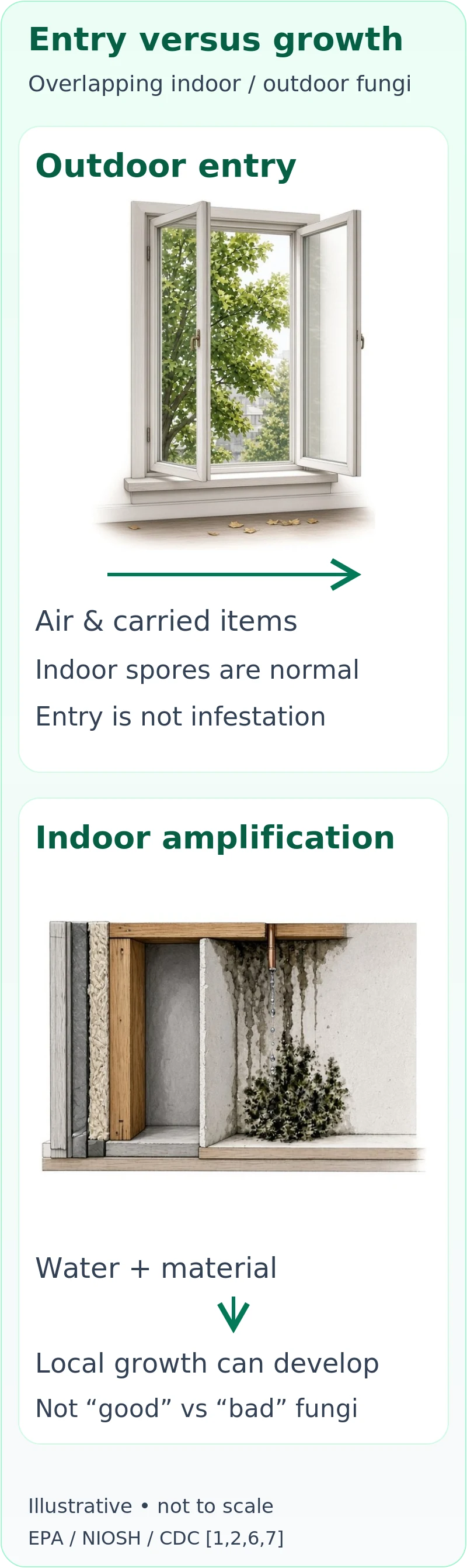
There are not two sealed categories of “bad indoor mold” and “good outdoor mold.” Outdoor fungi help break down organic material. Their spores enter buildings through doors, windows and ventilation, and on clothing, shoes and pets. Indoor and outdoor fungal populations overlap. [1, 2, 7]
What changes in a water-damaged building is the opportunity for indoor amplification: moisture and suitable materials allow fungi to grow inside rather than merely arrive from outdoors. Different fungi favor different moisture conditions and substrates. A persistently wet wall can support a different pattern of growth from a dry room receiving seasonal outdoor spores. Stachybotrys is one moisture-associated example, not a complete description of water-damaged-building exposure. [2, 3, 6]
That distinction matters when someone calls a species “outdoor mold” and dismisses an indoor finding. Outdoor origin does not mean a fungus cannot grow on damp indoor material. The reverse mistake is treating the name of a water-damage-associated fungus as proof that it caused a particular person's illness. The building history, location of growth, moisture and sampling method all matter. [2, 3, 5]
Water damage may also involve bacteria, their components, and chemicals released from building materials. NIOSH emphasizes that the specific agent responsible for an occupant's reaction may be uncertain. A species list can help an investigation, but it does not recreate that entire mixture or identify a single cause of symptoms. [2, 3]
How an environmental or functional clinician builds an exposure story
The starting point is not necessarily a test. A useful exposure history connects the building's story with the person's story: when a roof leaked, whether a room stayed damp, when symptoms began, whether a move or repair changed them, and what other health or life changes happened at the same time. The AWMF guideline recommends a detailed history and organ-focused assessment; functional and environmental-medicine writing places particular emphasis on the wider exposure context. [3, 14, 15]
Imagine someone who begins coughing after a basement floods. Finding the damp materials gives the building investigation a clear direction. If wheeze also develops, the clinical investigation might focus on asthma or allergy. If fatigue persists, that deserves evaluation too—but the sequence alone does not establish that a particular mycotoxin damaged mitochondria. An exposure story can be worth investigating before its mechanism is known. [3, 5, 7]
Following the basement example through—not a patient report
This is an illustrative scenario, not a reported case or a prediction of recovery. Suppose an assessor documents the leak and affected material, while a clinician evaluates the cough and wheeze. A functional assessment can explore the wider exposure history without waiting for a toxin result to address an identifiable airway problem or moisture source. [1, 3, 7]
Now imagine that a brief air sample reports few spores, a dust DNA report identifies selected fungi, and a urine panel flags a mycotoxin. These reports need not be measuring the same thing. The air result would not erase documented water damage; the DNA result would not demonstrate toxin production; and the urine finding would not identify the basement as its source. Ask what each sample represents and which decision it actually changes. [2, 3, 5, 9, 18]
The next step is to reconcile those questions—not select the most alarming report. The building plan should explain the source, repair and follow-up assessment; the clinical plan should track symptoms and function while considering other causes. Neither a particular score nor this example can promise a health outcome. [1, 2, 3]
Time away from a building can be informative, especially when a pattern repeats naturally. It is still not a controlled experiment: work schedules, other exposures and daily circumstances may change as well. Document what happens without deliberately returning to a suspect environment to provoke symptoms. NIOSH recommends discussing avoidance or alternative work locations with a healthcare provider when dampness-related concerns arise. [2, 3]
Exposure means a pathway, not just a name on a report
A functional assessment often asks how something could actually reach the person. Inhalation can involve spores, fragments and dust-associated material. Food provides a different route for mycotoxins; occupational dust can present circumstances quite different from everyday residential air. Skin contact is another possible route, particularly in some work settings. These pathways should be considered separately rather than added together into an unexplained “toxic load” number. [3, 4, 5, 18]
This is where building details become clinically relevant: a water-damaged material can be a source, disturbance can affect particle release, and the person's activities affect contact with it. But a complete hypothesis still needs evidence about what was present and what exposure occurred. The route, amount and duration matter. A positive dust or urine finding cannot supply all of those missing details. [3, 5, 18]
What effects on the body are established—and what remains disputed?
Damp, moldy environments are linked with upper-airway symptoms, cough, wheeze and worsening asthma in susceptible people. Mold exposure can cause allergy and irritation, including in some people without mold allergy. Hypersensitivity pneumonitis is a different immune-mediated lung condition that can occur in susceptible individuals; it is not simply another name for ordinary mold allergy. [1, 3, 7]
Selected people—especially those with weakened immune systems or certain chronic lung conditions—also face risks from fungal infection. Infection means fungal growth in the body; it is not interchangeable with inhaling spores, having a positive allergy test or detecting a mycotoxin. Appropriate evaluation depends on the person's risks and symptoms. [3, 7]
The debated claim is broader: that ordinary household inhalation of mycotoxins explains a chronic multisystem illness through a defined toxicity or CIRS mechanism. ACMT's 2025 position statement does not support indoor inhaled mycotoxins as an established systemic toxicant in these settings. AAAAI likewise describes insufficient evidence for attributing the range of nonspecific symptoms called toxic mold syndrome or CIRS to mycotoxin inhalation. [5, 12]
That is not a claim that a suffering person is imagining symptoms, or that a damp building is acceptable. It is a distinction between recognizing a problem and knowing its cause. The clinical guideline also discusses complex mixtures, extreme exposure situations and research gaps; routine residential exposure should not be assumed equivalent to heavily contaminated occupational or cleanup conditions. [3, 5]
What next: a practical route from concern to a plan
1. Document without disturbing. Record where and when the concern arose: leaks, odors, visible damage, photographs if safely obtainable, and symptom timing. Do not open a suspect wall simply to obtain a sample. [1, 2, 3]
2. Keep two evaluations moving. Ask an appropriate building professional about moisture and affected materials, and a healthcare professional about symptoms and medical risks. Neither assessment needs a consumer panel to answer every question before the other can begin. Urgent symptoms take priority over testing. [1, 3, 7]
3. Make testing earn its place. Before paying, ask what uncertainty the test resolves, what its limitations are, and what a positive or negative result would change. Request the complete report and the collection details, not just a score. [2, 3, 8, 18]
4. Ask for a repair-and-follow-up plan. Clarify the moisture source, what will be repaired or appropriately removed and cleaned, and how the work and any recurrence will be checked. A single number is not the whole completion assessment. For the building steps, see our safe mold remediation guide. Keep the clinical follow-up separate but informed by what the building investigation establishes. [1, 2, 3]
Building testing: start with the question, not the kit
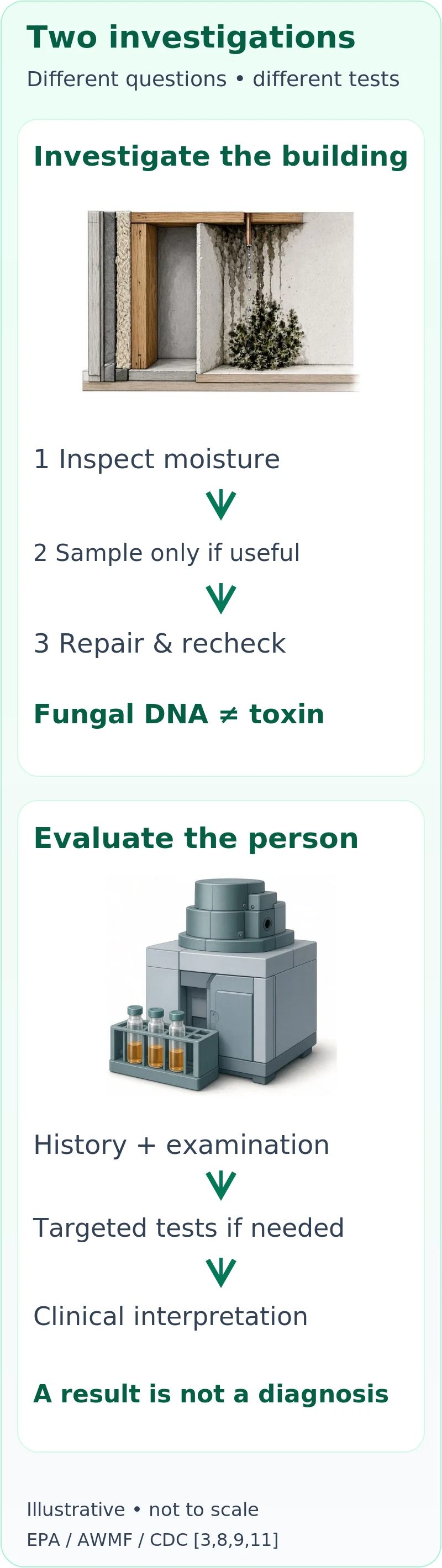
A building investigation asks where moisture and contamination are, how extensive they may be, and what needs correction. A medical evaluation asks what is happening to a person. The most expensive misunderstanding is buying one test and expecting it to answer both questions. [1, 2, 3]
Moisture inspection comes first
Start with leaks, condensation, staining, musty odors and the history of flooding or repairs. An experienced assessor may use moisture measurements and investigate concealed areas when the evidence warrants it. Hidden growth can occur behind walls, under flooring or above ceilings. Investigation should be planned carefully rather than beginning with indiscriminate demolition. [1, 2, 11]
If mold is already visible, EPA says sampling is usually unnecessary before addressing it. NIOSH finds thorough visual inspection and musty-odor assessment more reliable than routine short-term air sampling. That does not make every sample useless; it means a sample should resolve a defined uncertainty, not delay an obvious repair. [1, 2, 8]
Air samples and spore traps
An air sample describes what the method collected at that place and time. A short collection can miss an intermittent or concealed source; a negative result may not represent actual exposure. NIOSH cautions that spore counts and culture results do not capture the full range of exposures and cannot be converted into an individual's health risk. [2]
Microscopic spore counts and culture answer different questions. Microscopy can include viable and nonviable spores, whereas culture counts organisms that grow under the selected laboratory conditions. A low culture count is not evidence that all fungal material has disappeared. [3]
There are no universal health-based indoor mold thresholds that turn an ordinary air report into “safe for everyone” or “the cause of your illness.” EPA also has no federal airborne mold standard against which a report can certify compliance. Ask the assessor what the result changes in the building investigation. [2, 8]
Surface, tape, bulk-material and culture samples
Samples from a surface, a tape lift or a piece of material can help characterize a suspect location. Culture can help identify organisms that grow in the laboratory. These methods describe the sampled material—not all the material behind a wall, a person's inhaled dose or the medical cause of symptoms. EPA notes that surface sampling can sometimes help assess cleaning, when performed and interpreted appropriately. [3, 8]
DNA testing, qPCR and ERMI
Mold-specific quantitative PCR measures selected fungal DNA. EPA developed the Environmental Relative Moldiness Index, or ERMI, by analyzing indicator molds in settled dust and comparing homes within a research framework. It is not a direct mycotoxin assay or a medical diagnosis. A DNA measurement also is not, by itself, a demonstration that the detected fungus is currently growing. [3, 9]
Evidence and limits: EPA's fact sheet says ERMI has been peer reviewed for research purposes but not validated for non-research purposes, and EPA does not recommend routine public use. Research linking an index to health outcomes across groups does not establish an individual safe-occupancy threshold. Inspection for water damage remains central. ERMI is not a regulatory cleanup pass. [9]
Why an integrative practitioner may favor dust DNA—and what to clarify
A settled-dust test is attractive because it looks beyond a brief air collection and returns a selected fungal profile. CIRS practitioners may use that profile when discussing continued exposure, a move or re-entry after repairs. The rationale is to gather another clue about the environment, not merely to identify a patch visible on a wall. The ERMI research program and HERTSMI-2 developer guidance explain why dust and selected fungal indicators feature in these approaches. [9, 10, 15]
The collection plan still matters. A specimen from one room or one chosen surface cannot describe every cavity or floor of a building. Ask which areas it represents, why that collection method was selected, and how the assessor will reconcile the result with moisture findings. HERTSMI-2's own single-building-sample disclaimer is a useful reminder even within the model that promotes it. [1, 9, 10]
Different methods can disagree without one necessarily being fraudulent. A microscopy result describes collected structures; a culture describes what grew; qPCR describes selected DNA. None directly measures every fungal chemical. Before repeating tests, ask whether they are answering the same question on comparable material. A detailed assessment should explain a disagreement, not simply choose the most alarming number. [3, 5, 9]
MVOC air tests ask a different question
The MVOC section above explains why an odor-related chemical is not a specific marker of mold or mycotoxin dose. A combined report can contain several methods, but combining results does not make their meanings interchangeable. Ask what an MVOC measurement would change after inspection, rather than treating it as a medical diagnosis or a universal safe-occupancy test. [2, 3, 24]
Mycotoxin tests on dust or building material
These assays ask whether selected chemicals are detectable in the submitted sample. That is different from assuming toxin production based on a species name. But detecting a chemical on material does not establish how much reached an occupant, by which route, or whether it caused symptoms. A nondetection only addresses the substances and sample the method examined. [3, 5]
Evidence and limits: The clinical guideline does not recommend environmental mycotoxin measurements to diagnose indoor-mold illness, and says toxin testing is not necessary to decide that relevant mold contamination needs removal. A result may be a research measurement; that is not the same as a clinically interpretable illness test. [3]
Medical testing: matching a test to an actual clinical question
A useful appointment begins with symptoms, timing, medical history, medications, immune status and what is known about the building. Symptoms that seem worse in one location deserve documentation, but timing alone is not proof of a particular toxin. The clinician should consider mold-related conditions and other explanations rather than letting one environmental report dictate the diagnosis. [3, 5, 7]
Allergy, hypersensitivity pneumonitis and infection testing
For suspected mold allergy, an allergist may use a skin-prick test or mold-specific IgE blood testing alongside the history. A positive result indicates sensitization; it becomes evidence of a clinically relevant allergy when the symptoms and circumstances fit. Negative testing does not exclude every possible mold sensitization because available extracts and allergens are imperfect. [3]
Specific IgG testing can have a role in a specialist evaluation for hypersensitivity pneumonitis or other selected fungal conditions. It is not a general test for “mold toxicity,” proof of a particular building as the source, or evidence that a mycotoxin caused illness. IgE and IgG are immune measurements, not measurements of toxin dose. [3, 5]
When infection is a concern, clinicians may use imaging, cultures, molecular tests or tissue-based investigations suited to the suspected disease and the person's risk. A consumer urine mycotoxin panel does not substitute for that evaluation. Someone with a weakened immune system and new respiratory symptoms needs appropriate clinical advice, not reassurance from a negative environmental or urine result. [3, 7]
Urine and blood mycotoxin panels
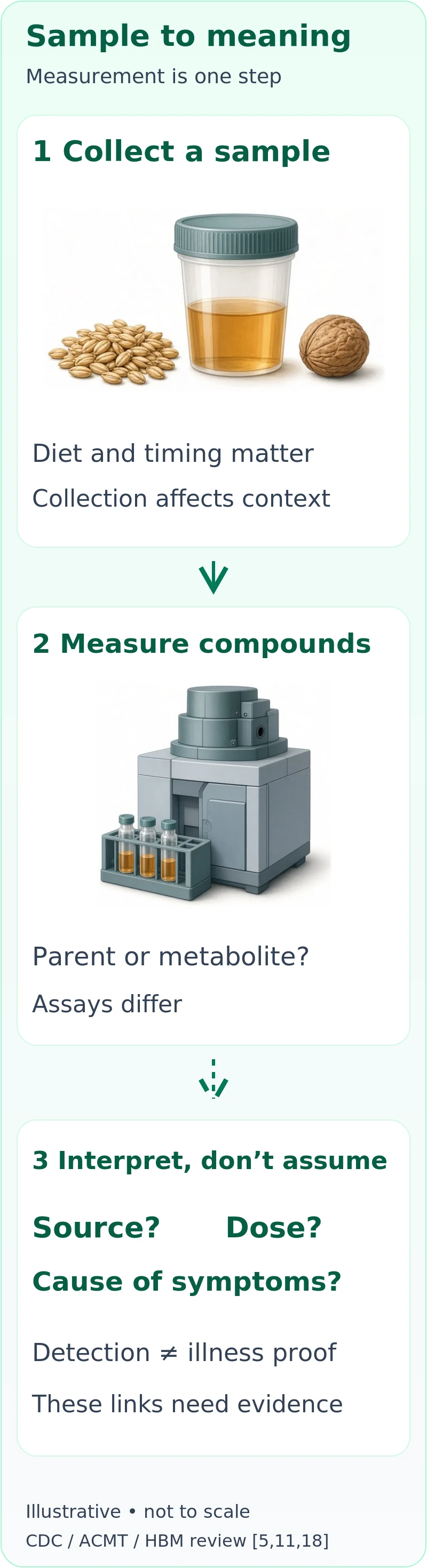
Laboratories can detect some mycotoxins or their metabolites in biological samples. The key distinction is analytical detection versus clinical validity: can the assay measure a substance, and does that measurement reliably diagnose the condition being proposed? Those are separate standards. [3, 5, 11]
CDC's 2015 report explains that low-level dietary exposure can lead to mycotoxins in healthy people's urine, and disease-predictive urine levels have not been established. ACMT's 2025 statement likewise emphasizes diet and cautions against attributing a urinary finding to a supposed earlier inhalation exposure. Blood detection does not automatically solve the source or clinical-meaning problem. [3, 5, 11]
A positive panel cannot, by itself, locate a building, distinguish food from indoor inhalation, or prove that the measured substance caused fatigue, brain fog or another symptom. A laboratory's flagged range is not automatically a validated threshold for household mold illness. A negative panel cannot clear a damp building or rule out mold allergy, irritation or other moisture-related problems. [3, 5, 11, 12]
For toxin-specific context, see aflatoxins and ochratoxin A above: a research exposure biomarker, a dietary risk assessment and a diagnosis of organ injury are not the same thing. The analyte and its biology matter more than a generic “toxin load” label. [18, 21, 23]
What a metabolite result actually means
A parent compound is the original chemical; a metabolite is a product of its processing. Some mycotoxin assays look for the parent, some for related metabolites, and some use preparation steps that change which forms can be measured. A glucuronide, for example, is a conjugated form made when a compound is linked to another chemical group during metabolism. The details are toxin- and method-specific—not evidence that every reported chemical behaves the same way. [18]
There is genuine science here. A major biomonitoring review distinguishes a measured substance from a validated exposure biomarker: to estimate intake, researchers need human data linking the measurement to exposure. Some mycotoxin biomarkers have useful research validation; others remain incompletely characterized. Even a validated exposure biomarker is not automatically a diagnostic test for a chronic symptom syndrome, and biomonitoring integrates sources rather than identifying a particular building. [18]
A laboratory may report concentration per volume of urine or adjust the result using creatinine, a substance excreted in urine. Dilution changes concentration. Hydration, kidney function and collection conditions can affect interpretation. Creatinine adjustment can reduce some dilution-related variation, but creatinine itself varies with factors including muscle mass and diet. It is not a mathematical conversion from a urine number into “how poisoned you are.” [18]
Timing matters too. A spot specimen samples a particular period; different toxins and metabolites have different absorption and elimination patterns. One measurement may reflect recent exposure better than longer-term exposure, and for some compounds human kinetic data remain limited. Sample handling, storage and laboratory preparation also matter. This is why a result needs its analyte name, units, collection details and method—not just a red or green marker. [18]
When comparing results over time, ask whether the same substances, units and collection conditions were used. A lower number could reflect a change in exposure, dilution, timing or method; it does not by itself prove successful treatment. A higher number is not automatically evidence that a treatment has “mobilized toxins.” That interpretation would require validation of the intervention and the clinical meaning of the change, not simply an appealing explanation. [3, 11, 18]
For the additional chemical examples, see macrocyclic trichothecenes, gliotoxin and derivatives, and zearalenone above. Identify whether a result describes a chemical family, a specific parent compound, or a named modified form before comparing its meaning with another result. The laboratory and specimen context still matters. [18, 30, 33, 36, 37]
Why functional and integrative practitioners use broader testing
Many readers arrive here after feeling unwell for a long time without a satisfying explanation. The appeal of a broader environmental evaluation is understandable: it connects a detailed exposure history, symptoms and laboratory findings instead of treating each complaint in isolation. Different integrative practitioners use different models; urine-panel approaches and the Shoemaker CIRS framework should not be treated as one uniform practice. [14, 15, 16]
The proposed biology: what practitioners mean by inflammation and impaired recovery
Functional and environmental-medicine authors often describe water-damaged-building illness as a problem of interacting exposures and host response rather than one toxin acting alone. Hope's review emphasizes inflammation, oxidative stress and differences in susceptibility. The Shoemaker model places persistent innate immune activation at its center. These frameworks overlap, but they are not identical, and neither makes every person's symptoms the same disease. [14, 15, 16]
Immune signaling: In the CIRS account, the body's early defense response fails to settle after exposure, and proposed changes in inflammatory or regulatory markers help explain continuing symptoms. In experimental toxicology, certain mycotoxins can alter immune-cell signaling. A 2025 systematic review of ochratoxin A describes changes in cytokines, macrophage function and other immune responses. Its studies include animal and cell models; a change in a laboratory system is not confirmation of persistent systemic immune dysfunction from a person's home. [15, 16, 19]
Oxidative stress: This describes an imbalance involving reactive molecules and protective systems, not a diagnosis that identifies an exposure source. Mold-illness authors propose that oxidative stress helps connect exposure with cellular symptoms. Experimental work supports the possibility that particular mycotoxins affect these processes under studied conditions. What remains unresolved is whether the same chain, at relevant doses and routes, explains an individual patient's ongoing illness. [14, 19]
Mitochondria and energy: Mitochondria help cells produce usable energy. Mycotoxin experiments have shown effects on mitochondrial function, and Brewer's patient study discusses mitochondrial dysfunction as a hypothesis for fatigue. It did not establish that a home's mold caused mitochondrial injury in the participating patients. Understanding the hypothesis helps explain why some clinicians focus on energy and antioxidant systems; it does not make fatigue a toxin-specific symptom or a urine panel a mitochondrial test. [13, 14, 19]
Barrier function: The gut lining and its junctions help regulate what passes between the intestinal contents and the body. Experimental studies reviewed in the ochratoxin A paper report altered barrier properties in intestinal cell systems and animals. This is relevant mechanistic research, particularly for ingestion, but it is not proof of a universal household-mold → “leaky gut” → chronic illness cascade in humans. The review itself emphasizes differences across experimental models and exposure conditions. [19]
These distinctions are useful rather than merely cautionary. They tell a clinician what should be measured and tested next: exposure, a relevant biological response, a meaningful clinical outcome, and whether changing exposure or care improves that outcome. A convincing account needs those links. It should also explain why another medical condition is less likely, rather than treating an interesting mechanism as the answer before the patient is evaluated. [3, 5, 18]
Urine panels as clues to an exposure hypothesis
Some practitioners interpret urine results as clues to mycotoxin exposure and use them within a model involving inflammation, oxidative stress or impaired clearance. Hope's 2013 review describes these proposed mechanisms and related clinical approaches. That explains the rationale; it does not establish that a urine level measures harmful retained “body burden,” identifies a home source or guides an effective treatment. [11, 12, 14]
There is published research behind the interest. A 2013 study by Brewer and colleagues tested patients with chronic fatigue syndrome and reported frequent urine detections. It used a previously studied healthy comparison group selected to have no known water-damaged-building exposure, rather than a newly matched group with the same exposure opportunities. The observational comparison therefore does not establish that household mycotoxins caused the illness or validate the panel for individual diagnosis. [13]
Evidence and limits: Human biomonitoring can be useful in exposure research, but the AWMF clinical guideline advises against using urine or blood mycotoxin biomonitoring to diagnose indoor-mold illness. A plausible biological story, an abnormal result and a patient's improvement can all generate hypotheses; they do not, together, establish diagnostic accuracy or prove a particular treatment worked. [3, 5, 11]
A related question is “provocation”—taking glutathione or using another intervention before collecting urine. This is not an established, validated diagnostic standard in the guidance cited here. A higher result after an intervention would not, by itself, resolve the source and disease-threshold problems. The cited guidance does not supply a validated glutathione-challenge interpretation. Do not start a challenge on your own to try to make a test positive. [3, 5, 11]
ERMI and HERTSMI-2 within the CIRS model
CIRS-focused practitioners often seek a measure of the building alongside a person's symptoms. Surviving Mold's HERTSMI-2 system uses DNA-based results for five selected fungi and proposes occupancy or re-entry categories for people treated within that model. Its own disclaimer says a single index cannot show every area of a building. [10, 15]
Evidence and limits: Those proposed categories are not EPA health thresholds or an independently established guarantee that a building is safe for a particular person. EPA's ERMI research caution still matters; a building score does not become a diagnostic test because it is considered together with symptoms. If a practitioner recommends testing, ask what decision it changes and what evidence validates that use. [3, 9, 10]
Inflammatory panels, HLA types and visual contrast sensitivity
The Shoemaker model describes patterns involving C4a, TGF-beta-1, MMP-9, MSH and other markers, along with selected HLA types and visual contrast sensitivity, or VCS. VCS measures the ability to distinguish contrast, rather than directly detecting mold or toxins. Practitioners use these findings to support a proposed pattern of persistent immune dysregulation. [15, 16]
Evidence and limits: These findings are not accepted definitive tests for indoor-mold illness. An abnormal inflammatory measurement or visual performance result is not specific proof of a mold cause; an HLA result does not demonstrate exposure or establish illness. The AWMF guideline specifically advises against cytokine measurements and VCS for this diagnostic purpose because evidence is insufficient. A supportive 2024 CIRS treatment review acknowledges that it was not a systematic review; its favorable interpretation does not independently validate the diagnostic model. [3, 12, 15, 16]
You do not need to choose between respectful care and honest uncertainty. It is reasonable to ask for a thorough history, attention to the building and consideration of other medical causes. It is also reasonable to ask a practitioner which findings would make them reconsider their explanation, and how they will avoid unnecessary tests or treatments.
Integrative care goals: what the model is trying to accomplish
An integrative plan is easiest to understand when its goals are separated: reduce a credible ongoing exposure, diagnose and treat identifiable conditions, support day-to-day function, and evaluate any additional proposed treatment on its own evidence. That approach allows a patient's environmental concerns to remain central without asking an unvalidated panel to direct every decision. Moisture correction and proper clinical assessment are common ground across the sources, even where explanations differ. [1, 3, 5, 14]
Address the environment without making the test the whole plan
For the building, the goal is not “zero fungal DNA.” Mold is naturally present in the environment. The goal is to correct moisture problems, remove relevant contamination safely, and check that the problem does not return. A proposed occupancy score can be discussed with the practitioner using it, but it should not replace a written explanation of the source, repair and follow-up assessment. [1, 2, 9, 10]
For the person, identified asthma, allergy or infection needs appropriate care. General symptoms and coexisting illnesses still deserve attention rather than being automatically absorbed into a mold diagnosis. Keeping a record of symptoms and function can make follow-up more useful, especially if multiple interventions are being considered. The question is not only whether a laboratory marker changed, but whether a meaningful health problem improved. [3, 5, 7]
Nutrition and recovery are not the same as a universal detox diet
Food is part of exposure assessment because it can be a source of mycotoxins. It is also the foundation of nutrition. WHO recommends sound food storage and a diverse diet to reduce risk; it does not turn the presence of a urinary metabolite into a reason to eliminate entire food groups. If someone is already restricting many foods or struggling to eat, a clinician and dietitian can help keep the plan practical and nutritionally adequate while genuine medical problems are investigated. [4, 11, 18]
What human treatment studies actually measured
Cholestyramine in a water-damaged-building study: A 2006 report combined a 26-patient time series with a double-blind, placebo-controlled trial completed by 13 participants. Seven were randomly assigned cholestyramine first and six placebo first. The authors reported improved symptom counts and visual contrast sensitivity during cholestyramine treatment, but not during the initial placebo period. The larger time series also changed exposure conditions; its before-and-after observations are not the same as randomized treatment effects. This is a genuine human study, but a very small, clinic-selected trial—not broad confirmation of a treatment protocol. Building samples were examined for fungi, but mycotoxins were not measured, so the study cannot identify a particular toxin as the treatment target. [25]
Oral glutathione in healthy adults: A six-month randomized, double-blind, placebo-controlled trial in 54 nonsmoking adults measured glutathione in blood and cells, with some additional immune measurements. The investigators reported increased glutathione stores. That supports a biological effect under the studied conditions; it was not a trial of symptom recovery, toxin removal or treatment of household-mold illness. Its population and outcomes differ from the cholestyramine study, so the two should not be presented as interchangeable proof of a “detox” approach. [26]
The distinction is not “research exists” versus “nothing exists.” Ask whether a study concerns the same people, exposure, treatment route and outcome as the claim being made. Animal studies of food-associated toxins and uncontrolled clinical observations can inform hypotheses without supplying the missing evidence for a residential-illness treatment. [3, 14, 23, 25, 26]
Why binders are discussed—and why supervision matters
CIRS and some integrative approaches use “binders,” including prescription cholestyramine, with the proposed goal of reducing reabsorption of substances from the gut. This rationale should be distinguished from proof that a particular binder removes the cause of a patient's symptoms. The placebo comparison is important, but the study’s size and scope still matter. The AWMF guideline considers detoxification therapy with cholestyramine insufficiently supported for indoor-mold illness. [3, 14, 25]
Cholestyramine does have an established pharmacologic action: it binds bile acids in the intestine. That is not the same as validation of a household-mycotoxin detox protocol. MedlinePlus lists constipation and other gastrointestinal side effects, and warns about interactions with other medicines. A prescriber and pharmacist need to review the whole medication and supplement list; this article does not provide a dose or timing regimen. Nonprescription charcoal or clay should not be assumed equivalent to a studied prescription treatment simply because all are called binders. [14, 20]
Glutathione and antioxidant approaches
Glutathione is part of the body's antioxidant systems. Environmental-medicine authors discuss supplementation as a way to support those systems. The rationale includes biochemical and experimental work; human studies need to be interpreted by the population, route and outcome they actually examined. That is a rationale for research and individualized discussion—not evidence that everyone with a positive urine panel needs glutathione or that supplementation establishes a diagnosis. [14, 26, 27]
The route of administration matters. In a randomized, double-blind crossover study of eight people with mild asthma, nebulized glutathione caused airway narrowing, with cough or breathlessness in some participants. This was an inhaled-route safety finding—not a test of oral supplementation or household-mold treatment. A substance being naturally present in the body does not make every formulation or route harmless. Care decisions should account for tolerability, existing illness and other medicines, and should not use symptom worsening as automatic proof that a detoxification process is working. No challenge, supplement or binder protocol is provided here. [14, 20, 27]
Agree what benefit and reassessment would look like
If an additional therapy is proposed, agree with the clinician on the intended symptom or functional benefit, who will supervise it, when to review progress, and which adverse effects should prompt contact or a change in the plan. Review medicines and supplements together. A plan should allow the explanation or treatment to be reconsidered—not treat every worsening symptom as proof that it is working. [3, 14, 20]
What should you do with a result?
Before ordering, ask: What specific question will this answer, and what will we do differently? A sample chosen to investigate a particular damp wall is different from a panel expected to explain every symptom. Ask what the method misses and whether the proposed cutoff predicts a clinical outcome or merely describes the laboratory's comparison range. [2, 3, 5, 8]
If you already have results, bring the complete report—not just a red flag or a score—to the relevant professional. For a building report, pair it with moisture findings and the sampling locations. For a medical report, pair it with your symptoms and history. Do not let a positive urine panel alone dictate costly remediation or a detox regimen; equally, do not let a low air count postpone fixing a documented leak and visible growth. [1, 2, 5, 11]
Our existing ERMI and HERTSMI-2 guide explores dust scores in more detail. The CIRS testing and treatment article explains that model without treating it as settled science. For next steps with the building, see safe mold remediation.
When professional care should not wait
Severe breathing difficulty, blue lips, or confusion with breathing trouble can signal an emergency: call 911 or your local emergency number rather than waiting for mold testing. If you have a weakened immune system or chronic lung disease, discuss new symptoms and mold exposure promptly with your treating team. Persistent or worsening symptoms also deserve evaluation even when the cause is uncertain. [3, 7, 17]
The bottom line is simpler than the testing marketplace can make it seem: spores are not toxins; color is not a diagnosis; and detection is not the same as disease. Address moisture and contamination safely, investigate symptoms properly, and ask every test to answer only the question it has evidence to answer. [1, 2, 3, 5]
Sources
Numbers in brackets in the text link to these sources. Sources were opened November 7–8, 2026, and the relevant passages were checked. Older research dates are shown so a historical study is not mistaken for current diagnostic validation. Developer and model-supporting sources are identified in the text.
- U.S. Environmental Protection Agency. A Brief Guide to Mold, Moisture and Your Home. Web guidance, updated February 18, 2026. https://www.epa.gov/mold/brief-guide-mold-moisture-and-your-home
- CDC / National Institute for Occupational Safety and Health. Mold, Testing, and Remediation. Web guidance, updated October 28, 2026. https://www.cdc.gov/niosh/mold/testing-remediation/index.html
- Hurraß J, Heinzow B, Walser-Reichenbach S, et al. AWMF mold guideline “Medical clinical diagnostics for indoor mold exposure” — Update 2023, AWMF Register No. 161/001. Allergologie Select. 2024;8:90–198. doi:10.5414/ALX02444E. Full text: PubMed Central; archived XML: Europe PMC. https://pmc.ncbi.nlm.nih.gov/articles/PMC11097193/
- World Health Organization. Mycotoxins. Fact sheet, October 2, 2023. https://www.who.int/news-room/fact-sheets/detail/mycotoxins
- Leikin J, Holland MG, Kurt TL, McKay CA, Stolbach AI. ACMT Position Statement: Medical Toxicology Considerations in the Diagnosis and Treatment of Patients with Concerns About Mold-Related Inhalation Exposures. American College of Medical Toxicology. August 13, 2025. https://www.acmt.net/news/acmt-position-statement-medical-toxicology-considerations-in-thediagnosis-and-treatment-of-patients-with-concerns-aboutmold-related-inhalation-exposures/
- Centers for Disease Control and Prevention. Facts About Stachybotrys chartarum. Web guidance. https://www.cdc.gov/mold-health/data-research/facts-stats/index.html
- Centers for Disease Control and Prevention. Mold. Web guidance. https://www.cdc.gov/mold-health/about/index.html
- U.S. Environmental Protection Agency. Mold Testing or Sampling. Web guidance, updated January 14, 2026. https://www.epa.gov/mold/mold-testing-or-sampling
- U.S. Environmental Protection Agency. The Environmental Relative Moldiness Index (ERMI). Fact sheet, September 2021; current EPA ERMI webpage also opened (updated September 29, 2026). https://www.epa.gov/system/files/documents/2021-09/updated-fact-sheet.ermi-_9.9.21.final_new-template_508-compliant_0.pdf
- Surviving Mold. HERTSMI-2 Scoring System. Undated developer guidance. https://www.survivingmold.com/resources-for-patients/diagnosis/hertsmi-2
- Kawamoto M, Page E. Notes from the Field: Use of Unvalidated Urine Mycotoxin Tests for the Clinical Diagnosis of Illness — United States, 2014. MMWR Morb Mortal Wkly Rep. 2015;64(6):157–158. https://www.cdc.gov/mmwr/preview/mmwrhtml/mm6406a7.htm
- American Academy of Allergy, Asthma & Immunology. Toxic Mold Syndrome: Separating Fact from Fiction. Patient education webpage, dated July 29, 2026. https://www.aaaai.org/tools-for-the-public/conditions-library/allergies/toxic-mold
- Brewer JH, Thrasher JD, Straus DC, Madison RA, Hooper D. Detection of Mycotoxins in Patients with Chronic Fatigue Syndrome. Toxins. 2013;5(4):605–617. doi:10.3390/toxins5040605. Full text: PubMed Central; archived XML: Europe PMC. https://pmc.ncbi.nlm.nih.gov/articles/PMC3705282/
- Hope J. A Review of the Mechanism of Injury and Treatment Approaches for Illness Resulting from Exposure to Water-Damaged Buildings, Mold, and Mycotoxins. The Scientific World Journal. 2013;2013:767482. doi:10.1155/2013/767482. Full text: PubMed Central; archived XML: Europe PMC. https://pmc.ncbi.nlm.nih.gov/articles/PMC3654247/
- Shoemaker R, Johnson K, Jim L, et al. Diagnostic Process for Chronic Inflammatory Response Syndrome (CIRS): A Consensus Statement. Report of the Consensus Committee of Surviving Mold. Internal Medicine Review. 2018;4(5). https://www.survivingmold.com/Publications/CIRS_diagnostic_protocol_final_5_1_2018.pdf
- Dooley M, Vukelic A, Jim L. Chronic inflammatory response syndrome: a review of the evidence of clinical efficacy of treatment. Annals of Medicine and Surgery. 2024;86(12):7248–7254. doi:10.1097/MS9.0000000000002718. Full text: PubMed Central; archived XML: Europe PMC. https://pmc.ncbi.nlm.nih.gov/articles/PMC11623837/
- National Heart, Lung, and Blood Institute. Respiratory Failure: Symptoms. Web guidance, updated March 24, 2022. https://www.nhlbi.nih.gov/health/respiratory-failure/symptoms
- Cramer B, Visintin L, Maris E, et al. Human biomonitoring of mycotoxins: key challenges and future directions. Mycotoxin Research. 2026;42:13. Published online December 23, 2025. doi:10.1007/s12550-025-00612-2. Full text: PubMed Central; archived XML: Europe PMC. https://pmc.ncbi.nlm.nih.gov/articles/PMC12727801/
- Mubarik Y, Boyetey ST, Aikins AR, Mutocheluh M. Effect of Ochratoxin A (OTA) on the Immune System: A Systematic Review. Toxins. 2025;17(5):256. doi:10.3390/toxins17050256. Full text: PubMed Central; archived XML: Europe PMC. https://pmc.ncbi.nlm.nih.gov/articles/PMC12115609/
- MedlinePlus / American Society of Health-System Pharmacists. Cholestyramine Resin: Drug Information. Revised March 15, 2026. https://medlineplus.gov/druginfo/meds/a682672.html
- National Toxicology Program, U.S. Department of Health and Human Services. Aflatoxins. Report on Carcinogens, Fifteenth Edition. 2021. https://ntp.niehs.nih.gov/sites/default/files/ntp/roc/content/profiles/aflatoxins.pdf
- International Agency for Research on Cancer. List of Classifications, Volumes 1–142. Current public table checked November 7, 2026; table last updated July 23, 2026. Aflatoxins: Group 1; ochratoxin A: Group 2B. https://monographs.iarc.who.int/list-of-classifications/
- EFSA Panel on Contaminants in the Food Chain (CONTAM), Schrenk D, et al. Risk assessment of ochratoxin A in food. EFSA Journal. 2020;18(5):6113. doi:10.2903/j.efsa.2020.6113. Full text: PubMed Central; archived XML: Europe PMC. https://pmc.ncbi.nlm.nih.gov/articles/PMC10464718/
- U.S. Environmental Protection Agency. What does mold smell like? Web guidance, updated September 8, 2026. https://www.epa.gov/mold/what-does-mold-smell
- Shoemaker RC, House DE. Sick building syndrome (SBS) and exposure to water-damaged buildings: time series study, clinical trial and mechanisms. Neurotoxicology and Teratology. 2006;28(5):573–588. doi:10.1016/j.ntt.2006.07.003. Full publisher-formatted author copy opened. https://www.survivingmold.com/docs/Resources/Shoemaker%20Papers/NTT5863.pdf
- Richie JP Jr, Nichenametla S, Neidig W, et al. Randomized controlled trial of oral glutathione supplementation on body stores of glutathione. European Journal of Nutrition. 2015;54(2):251–263. doi:10.1007/s00394-014-0706-z. Primary study abstract opened through the authors’ Penn State record and Europe PMC. https://pure.psu.edu/en/publications/randomized-controlled-trial-of-oral-glutathione-supplementation-o/
- Marrades RM, Roca J, Barberà JA, de Jover L, MacNee W, Rodriguez-Roisin R. Nebulized glutathione induces bronchoconstriction in patients with mild asthma. American Journal of Respiratory and Critical Care Medicine. 1997;156(2 Pt 1):425–430. doi:10.1164/ajrccm.156.2.9611001. Primary study abstract opened via Europe PMC; PubMed record linked. https://pubmed.ncbi.nlm.nih.gov/9279219/
- U.S. Environmental Protection Agency. Mold Course Chapter 1: Introduction to Molds. Lessons 1–2 and 4–6; updated February 18, 2026. https://www.epa.gov/mold/mold-course-chapter-1
- Brandt M, Brown C, Burkhart J, et al. Mold Prevention Strategies and Possible Health Effects in the Aftermath of Hurricanes and Major Floods. CDC. MMWR Recommendations and Reports. 2006;55(RR-8). Sections: Mold Biology and Factors That Produce Mold Growth. https://www.cdc.gov/mmwr/preview/mmwrhtml/rr5508a1.htm
- Zhu M, Cen Y, Ye W, Li S, Zhang W. Recent Advances on Macrocyclic Trichothecenes, Their Bioactivities and Biosynthetic Pathway. Toxins. 2020;12(6):417. doi:10.3390/toxins12060417. Full text read via Europe PMC. https://pmc.ncbi.nlm.nih.gov/articles/PMC7354583/
- Došen I, Andersen B, Phippen CBW, Clausen G, Nielsen KF. Stachybotrys mycotoxins: from culture extracts to dust samples. Analytical and Bioanalytical Chemistry. 2016;408:5513–5526. doi:10.1007/s00216-016-9649-y. Primary publisher full text opened; analytical building case study, not clinical causal validation. https://link.springer.com/article/10.1007/s00216-016-9649-y
- Cundliffe E, Cannon M, Davies J. Mechanism of inhibition of eukaryotic protein synthesis by trichothecene fungal toxins. Proceedings of the National Academy of Sciences USA. 1974. doi:10.1073/pnas.71.1.30. Published primary abstract read via Europe PMC; PubMed record linked. https://pubmed.ncbi.nlm.nih.gov/4521056/
- Vidal-García M, Redrado S, Domingo MP, et al. Production of the Invasive Aspergillosis Biomarker Bis(methylthio)gliotoxin Within the Genus Aspergillus: In Vitro and in Vivo Metabolite Quantification and Genomic Analysis. Frontiers in Microbiology. 2018;9:1246. doi:10.3389/fmicb.2018.01246. Primary publisher full text read. https://www.frontiersin.org/journals/microbiology/articles/10.3389/fmicb.2018.01246/full
- Mercier T, Reséndiz Sharpe A, Waumans D, Desmet K, Lagrou K, Maertens J. Gliotoxin and bis(methylthio)gliotoxin are not reliable as biomarkers of invasive aspergillosis. Mycoses. 2019;62(10). doi:10.1111/myc.12967. Published primary abstract read via Europe PMC; PubMed record linked. https://pubmed.ncbi.nlm.nih.gov/31313395/
- Johannessen LN, Nilsen AM, Løvik M. The mycotoxins citrinin and gliotoxin differentially affect production of the pro-inflammatory cytokines tumour necrosis factor-alpha and interleukin-6, and the anti-inflammatory cytokine interleukin-10. Clinical & Experimental Allergy. 2005. doi:10.1111/j.1365-2222.2005.02249.x. Published primary abstract read via Europe PMC; cell-line experiment, not a clinical trial. https://pubmed.ncbi.nlm.nih.gov/15969670/
- Eriksen GS, Pennington J, Schlatter J, et al. Zearalenone. In: Safety Evaluation of Certain Food Additives and Contaminants. WHO Food Additives Series 44. Joint FAO/WHO Expert Committee on Food Additives. Geneva: World Health Organization; 2000. Full monograph read in relevant sections; historical evaluation, no numerical guidance value reproduced. https://www.inchem.org/documents/jecfa/jecmono/v44jec14.htm
- EFSA Panel on Contaminants in the Food Chain (CONTAM), Knutsen HK, et al. Risks for animal health related to the presence of zearalenone and its modified forms in feed. EFSA Journal. 2017;15(7):4851. doi:10.2903/j.efsa.2017.4851. Published opinion abstract read via Europe PMC; animal-feed assessment, not a human clinical study. https://pubmed.ncbi.nlm.nih.gov/32625539/